AI-Enhanced Vision: How FUJIFILM Technology Helps Doctors Detect Disease Earlier
In modern healthcare facilities around the world, radiologists face a mounting challenge: analysing an ever-increasing volume of complex medical images with the precision and speed patients deserve. Each scan contains countless details that could signal the difference between health and disease, early intervention and late-stage treatment. In this high-stakes environment, FUJIFILM innovation is creating a remarkable partnership between human expertise and artificial intelligence.
The Radiologist’s Challenge
Consider the daily reality for radiologists: hundreds of images to interpret, each potentially containing subtle indicators of serious conditions. A small lung nodule that might be early-stage cancer. Faint patterns suggesting interstitial lung disease. A nearly imperceptible pneumothorax (collapsed lung) requiring immediate intervention.
These professionals are highly trained to detect such anomalies, but the human eye and brain—even those of experts—face limitations when processing vast amounts of visual data under time constraints. This challenge is where FUJIFILM technology impact is most profoundly felt in modern healthcare.
REiLI: The Intelligent Assistant
FUJIFILM’s REiLI platform represents a significant leap forward in medical imaging AI. Named after the Japanese word “reiri” meaning “smart” or “clever,” this AI assistant exemplifies how FUJIFILM beyond cameras is transforming healthcare through digital innovation.
“REiLI doesn’t replace radiologists—it enhances their capabilities,” explains Dr. Sarah Chen, a thoracic radiologist at a major urban medical center. “The AI helps prioritize cases and flags subtle findings I need to focus on. It’s an invaluable tool that enhances our diagnostic confidence and efficiency.”
This collaboration between human expertise and artificial intelligence creates a powerful synergy. The AI excels at consistent pattern recognition across thousands of images, while radiologists contribute their clinical judgment, contextual understanding, and decision-making expertise.
How REiLI Works: AI in Action
REiLI’s capabilities span multiple imaging modalities and clinical applications:
Comprehensive Analysis Across Imaging Types
- Chest X-ray interpretation – Detecting and highlighting potential abnormalities like nodules, pneumonia, and pneumothorax
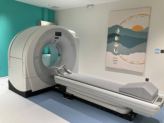
- CT scan analysis – Identifying and characterizing pulmonary nodules, emphysema patterns, and other thoracic abnormalities
- Mammography support – Assisting in the detection of potential breast cancer indicators
- MRI evaluation – Helping identify regions of interest in neurological, musculoskeletal, and other specialised imaging
Specific Clinical Applications
REiLI’s specialized algorithms target numerous conditions, including:
- Pneumothorax detection
Rapidly identifying potentially life-threatening collapsed lungs - Interstitial lung disease patterns
Recognizing the subtle patterns indicative of various lung diseases - Pulmonary nodule detection and characterization –
Finding and providing initial assessment of potential lung cancers - Bone fracture identification
Highlighting areas of potential breaks that might be overlooked - Cerebral hemorrhage detection
Assisting in rapid identification of brain bleeds in emergency settings
The system operates seamlessly within existing radiology workflows, integrating with FUJIFILM’s SYNAPSE PACS (Picture Archiving and Communication System) to provide radiologists with AI insights directly within their regular working environment.
The Technology Behind the AI
REiLI’s impressive capabilities stem from FUJIFILM’s unique combination of imaging expertise and AI development:
Decades of Image Processing Excellence
Long before AI became a healthcare buzzword, FUJIFILM had established itself as a leader in image processing. The company’s experience in enhancing visual clarity, extracting meaningful data from images, and optimizing visualisation provided the foundation for its medical AI development.
“What makes REiLI different is that it comes from a company with deep imaging expertise,” notes an imaging informatics specialist at a university hospital system. “FUJIFILM understands both the technical aspects of medical imaging and the clinical workflow needs. That perspective shapes how their AI integrates into real-world practice.”
Advanced Machine Learning Techniques
REiLI employs sophisticated deep learning algorithms trained on diverse, carefully-curated datasets. These models continually improve through ongoing training and validation, ensuring they perform reliably across different patient populations and clinical settings.
Importantly, the system is designed to maintain appropriate human oversight. REiLI provides suggestions and highlights areas of interest, but diagnostic decisions remain firmly in the hands of qualified healthcare professionals.
Measured Impact on Care
The integration of REiLI into clinical practice has yielded measurable benefits:
Enhanced Efficiency
Studies of AI assistance in radiology consistently show improvements in reading time efficiency. Radiologists using REiLI can prioritise their workload more effectively, focusing their expertise where it’s most needed while allowing the AI to help with initial screening and triage.
“During busy shifts, having REiLI help prioritize cases with potential critical findings allows us to address urgent situations faster,” explains a radiologist who uses the system daily. “This can make a real difference in time-sensitive situations like potential stroke or pneumothorax.”
Improved Diagnostic Accuracy
Research suggests that the combination of human expertise and AI assistance leads to better outcomes than either alone. In particular, AI support has shown promise in:
- Reducing missed findings in complex images
- Providing consistent assessment of features like nodule size and growth over time
- Maintaining diagnostic quality during high-volume reading sessions when human fatigue might be a factor
Democratising Expertise
Perhaps most significantly, REiLI helps extend radiological expertise to settings where specialist resources are limited. In regions facing radiologist shortages or in smaller hospitals without subspecialists, AI assistance can help general radiologists provide higher levels of care.
“In our regional network, REiLI helps our general radiologists feel more confident when specialized expertise isn’t immediately available,” notes a healthcare administrator from a rural hospital system. “It’s like having a second opinion built into your workflow.”
The Human Element Remains Central
Despite these technological advances, FUJIFILM’s approach keeps healthcare professionals at the center of the diagnostic process. REiLI is designed as an assistant rather than a replacement—augmenting human capabilities rather than attempting to automate them away.
This philosophy reflects an understanding that medical diagnosis isn’t merely pattern recognition. It requires integrating visual findings with clinical history, laboratory results, and the nuanced judgment that comes from years of training and experience.
“What I appreciate about the system is that it respects my role as a physician,” says a radiologist who was initially skeptical about AI. “It doesn’t try to make decisions for me—it gives me tools that enhance my ability to make better decisions for my patients.”
Experiencing AI Innovation Firsthand
For healthcare professionals and administrators interested in experiencing these AI capabilities firsthand, the FUJIFILM Technology Centre Dubai offers an immersive look at how REiLI integrates into the diagnostic workflow. Visitors can see demonstrations of the technology across various imaging modalities and understand how it complements FUJIFILM’s comprehensive medical imaging ecosystem.
“Visitors to our FTC Experience Dubai are often surprised by how seamlessly the AI integrates into the radiologist’s workflow,” explains a FUJIFILM healthcare specialist. “They can observe how REiLI highlights findings directly within the SYNAPSE viewing environment, making the AI insights immediately actionable.”
The Future of AI-Enhanced Diagnostics
As healthcare continues to digitise and imaging volumes grow, the partnership between radiologists and AI assistants like REiLI will become increasingly important. FUJIFILM continues to expand REiLI’s capabilities, developing new algorithms and refining existing ones to address more clinical scenarios.
The company’s vision extends beyond individual AI applications to creating an integrated ecosystem where diagnostic AI works harmoniously with imaging hardware, healthcare IT systems, and clinical workflows. This holistic approach reflects FUJIFILM’s societal impact mission to improve healthcare through comprehensive technology solutions.
For patients, this progression means more consistent, accurate, and timely diagnoses. For healthcare systems, it offers a path to maintain diagnostic quality while managing increasing imaging volumes. And for radiologists, it provides tools that enhance their expertise rather than attempting to replace it.
To explore how FUJIFILM’s REiLI platform could enhance diagnostic capabilities in your healthcare facility, consider arranging a visit to the FUJIFILM Technology Centre Dubai. Here, you can experience firsthand the impact of FUJIFILM technology on daily life through its advanced medical applications, and see why the question “What does FUJIFILM make besides cameras?” has such a profound answer in the healthcare domain.
Through REiLI and its broader healthcare solutions, FUJIFILM continues to demonstrate how a legacy of imaging excellence can translate into technologies that help physicians see more clearly—and help patients heal faster through earlier, more accurate diagnosis.
For more on FUJIFILM’s Radiology technology, visit fujifilm.com/ae.